Virological
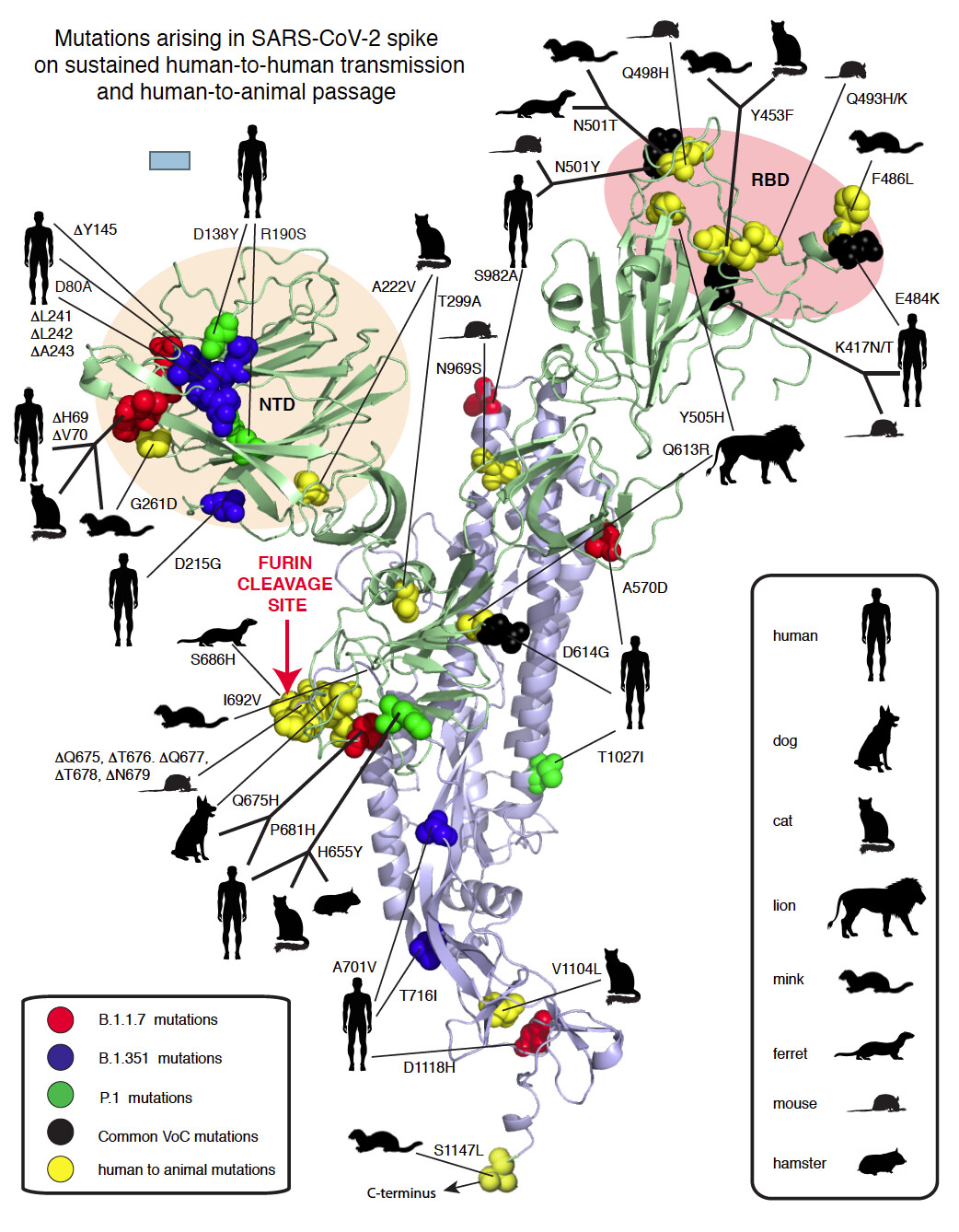
Mutations arising in SARS-CoV-2 spike on sustained human-to-human transmission and human-to-animal passage
SARS-CoV-2 coronavirus
rfgarry
January 18, 2021, 3:35pm
5
image
1042×1338 389 KB
show post in topic