Primer erosion analysis for two Andes virus Real time RT-PCR systems
Manfred Weidmann 1,2, Nicole Tischler3, Frank T. Hufert1, Idrissa Dieng4
1 Brandenburg Medical School Theodor Fontane, Institute of Microbiology and Virology, Senftenberg, Germany
2Institute of Animal Hygiene and Veterinary Public Health, University Leipzig, Germany
3 Molecular Virology Laboratory, Fundación Ciencia, Santiago, Chile
4 Institut Pasteur de Dakar, Senegal
We analysed if the oligonucleotides of existing Andes virus (Orthohantavirus andesense, ANDV) real time RT-qPCR fit to the current ANDV outbreak genome sequences. In project FhG InSan I 0301-V-4303 in the early 2000 a real time qRT-PCR for the detection of the S-segment of ANDV was developed. The S-segment of ANDV was ligated into pCRII and an in vitro RNA standard derived as described (1). At the time the analytical sensitivity of the assay was shown to be 10 RNA molecules/reaction using the transcribed in vitro RNA standard. The assay was shown not to cross detect viral RNA of Sin Nombre virus.
In 2007 Kramski and Colleagues developed an RT-qPCR for rapid detection of ANDV targeting the S gene (2). This system also had an analytical sensitivity of 10 RNA molecules/reaction and high technical reproducibility confirming his usefulness for ANDV specific detection using different platforms (3).
Table 1. Details of oligonucleotides analysed
For the analysis we used the published kmer screen based Covidmutants tool (4) and an in-house pipeline from IPD namely O.Mi.Count (Oligo Mismatches Counter).
Results of Analysis with Covidmutants tool
Table 2. Results of primer screen
* NCBIvirus download of 183 ANDV S-segment sequences on 12.05.2026 including one new sequence of the current outbreak (PZ385163.1 isolate ANDV/Switzerland/Hu-3337/2026)
** All ANDV S-segment sequences downloaded from NCBI were sorted by size, aligned to the primers of the amplicon and fragments that did not fully align with the amplicon were removed.
The results indicate that the shorter 68nt amplicon amplified by primers & probe 1 cover a larger variety of reported ANDV S-segment sequences than the 199 nt amplicon defined by primers & probe 2 including the current new ANDV sequence (see figure 2).
Results of Analysis with O.Mi.Count
For primers & probe 1 only 3 sequences shows mutations on binding sites (Figure 1A). The analysis of primers & probe 2 shows that among all analysed sequences (n = 97), 26 exhibit mutations in oligonucleotide sequence targets (Figure 1B). A single mutation is located in 14 sequences from Argentina (n = 13) and the new sequence from Switzerland (n = 1) in the forward primer but not at the critical 3’ end previously described to be detrimental (Figure S1 / Figure S2)(5).
Despite genetic evolution, ANDV has not escaped detection by existing RT-qPCR systems. Routine surveillance is recommended, but urgent assay redesign is not required. This in silico analysis provides reassuring evidence that current ANDV RT-qPCR assays remain compatible with re-emerged strains. The single shared C→T mutation at position 16 of primers & probe 2 is unlikely to compromise diagnostic sensitivity.
- Figure 1A
- Figure 1B
Figure 1: Mutation analysis by O.Mi.Count: Figure 1A primers & probe 1, Figure 1B: primers & probe 2.
Direct alignment of Primers & Probes
In direct alignment primers & probe 1 and 2 both align to the ANDV S-segment sequences derived from patient material of the ongoing outbreak and should both be able to amplify these current S-segment targets. Differences to the me ANDV S-segment sequence are highlighted by colours.
Primer 1
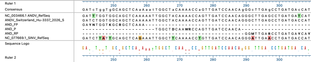
Primer 2

Figure 2: Primer1: Direct alignment of primers & probe 1, Primer 2 alignment of primers & probe
Conclusion
The analysis has provided in silico evidence that both primer sets should be able to amplify the ANDV S-segment targets of the current outbreak.
Acknowledgments
We would like to thanks Walter Zingg the clinician from Zurich and the teams from University of Zurich and Geneva University Hospitals who generated and made available the sequence of the ANDV strain detected in Swizerland.This work was an activity in the European Union’s Horizon Europe research and innovation programme under grant agreement No. 101137132 (PREPARE TID).
Contact :
Dr Idrissa Dieng ([email protected])
References
1. Weidmann M, Rudaz V, Nunes MR, Vasconcelos PF, Hufert FT. Rapid detection of human pathogenic orthobunyaviruses. J Clin Microbiol. 2003;41(7):3299-305.
2. Vial C, Martinez-Valdebenito C, Rios S, Martinez J, Vial PA, Ferres M, et al. Molecular method for the detection of Andes hantavirus infection: validation for clinical diagnostics. Diagn Microbiol Infect Dis. 2016;84(1):36-9.
3. Kramski M, Meisel H, Klempa B, Kruger DH, Pauli G, Nitsche A. Detection and typing of human pathogenic hantaviruses by real-time reverse transcription-PCR and pyrosequencing. Clin Chem. 2007;53(11):1899-905.
4. Weidmann M, Graf E, Lichterfeld D, Abd El Wahed A, Bekaert M. Efficient Screening of Long Oligonucleotides Against Hundred Thousands of SARS-CoV-2 Genome Sequences. Frontiers in Virology. 2022;Volume 2 - 2022.
5. Stadhouders R, Pas SD, Anber J, Voermans J, Mes TH, Schutten M. The effect of primer-template mismatches on the detection and quantification of nucleic acids using the 5’ nuclease assay. J Mol Diagn. 2010;12(1):109-17.
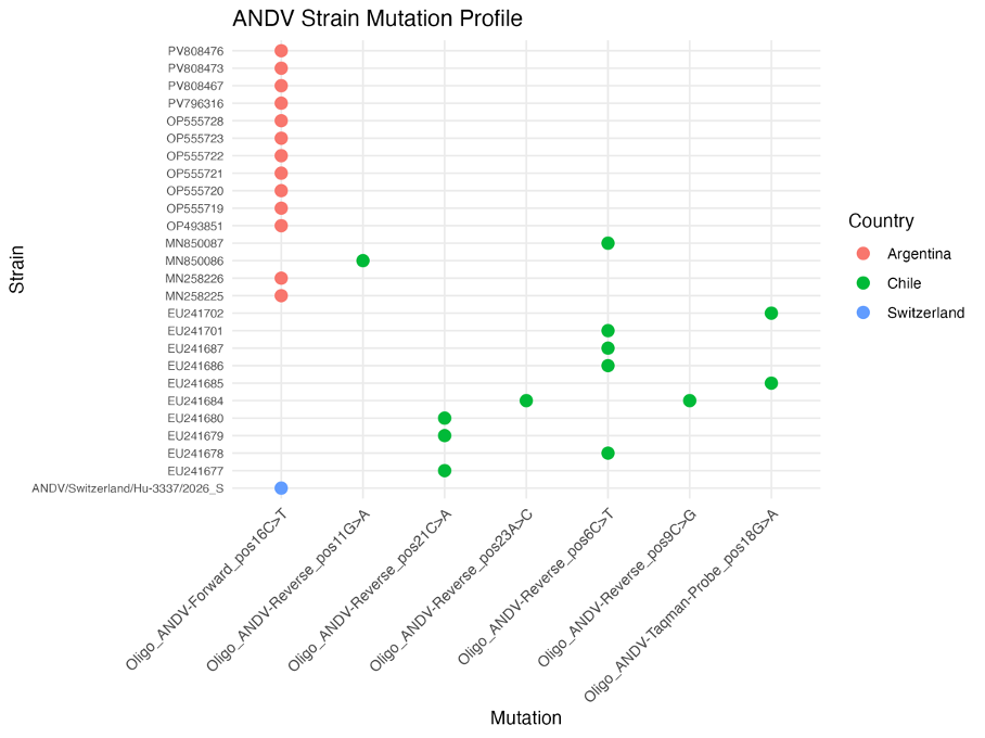
Figure S1. Distribution of mutations observed in primer & probe 2 across strains.
Figure S2. Geolocation of changed strains carrying the mutations in Figure 1.