2018 LASV sequencing, continued

Following up on our first post on the sequencing efforts by the collaboration between the Irrua specialist Teaching Hospital (ISTH), Institute for Lassa Fever Research and Control (ILFRC), Irrua, Edo State, Nigeria, The Bernhard-Nocht Institute for Tropical Medicine (BNITM), Hamburg, Germany, Public Health England (PHE), we now make a available sequences from 8 additional 2018 samples, amounting to a total of 15 samples:

https://github.com/ISTH-BNITM-PHE/LASVsequencing

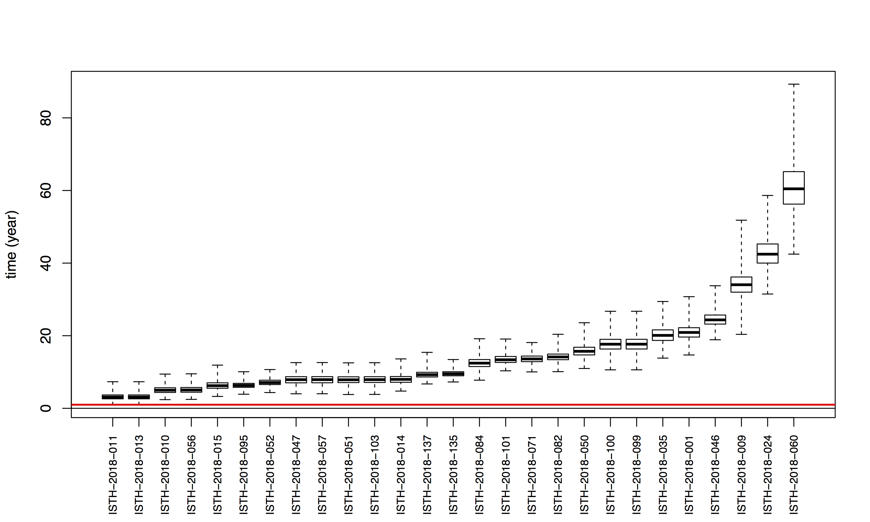
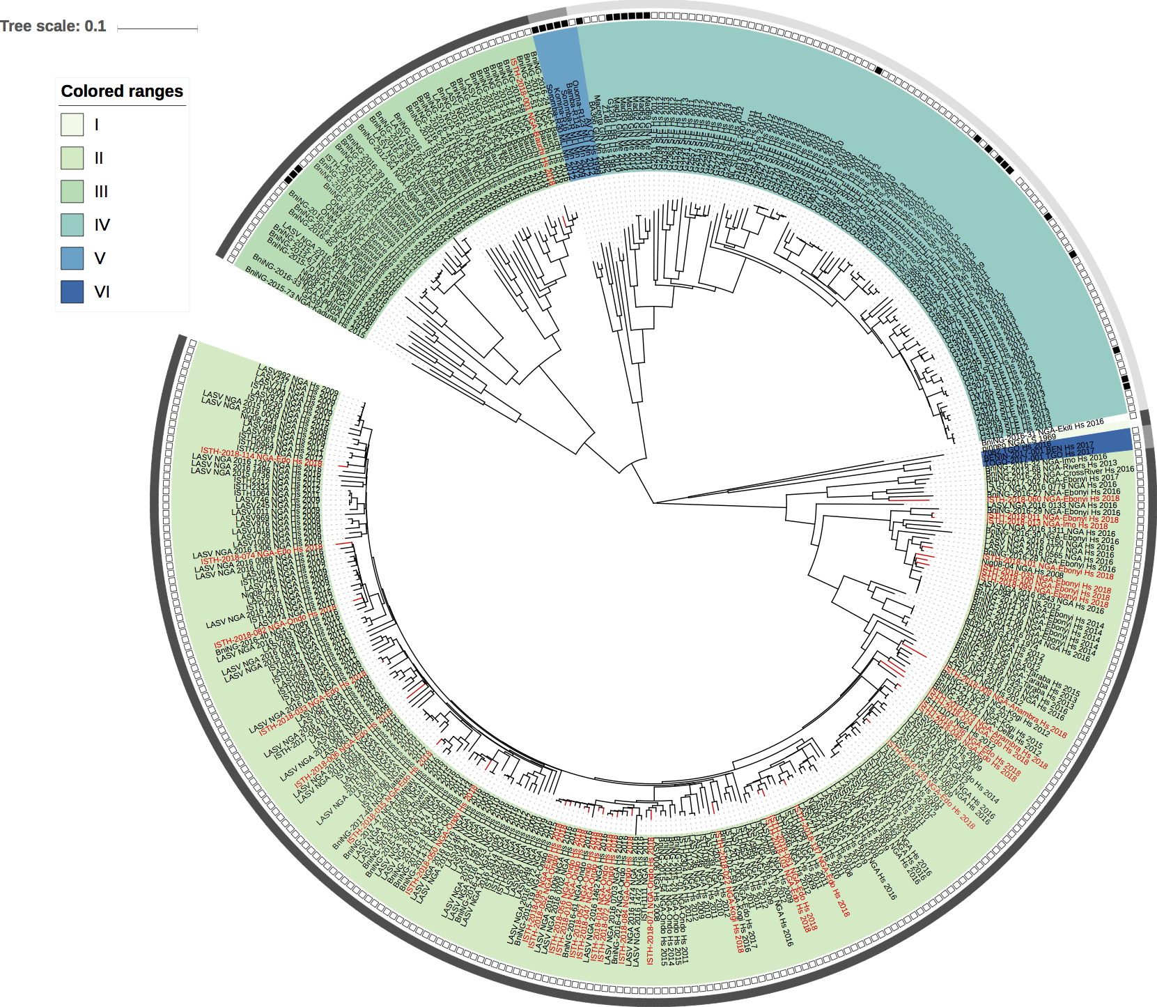
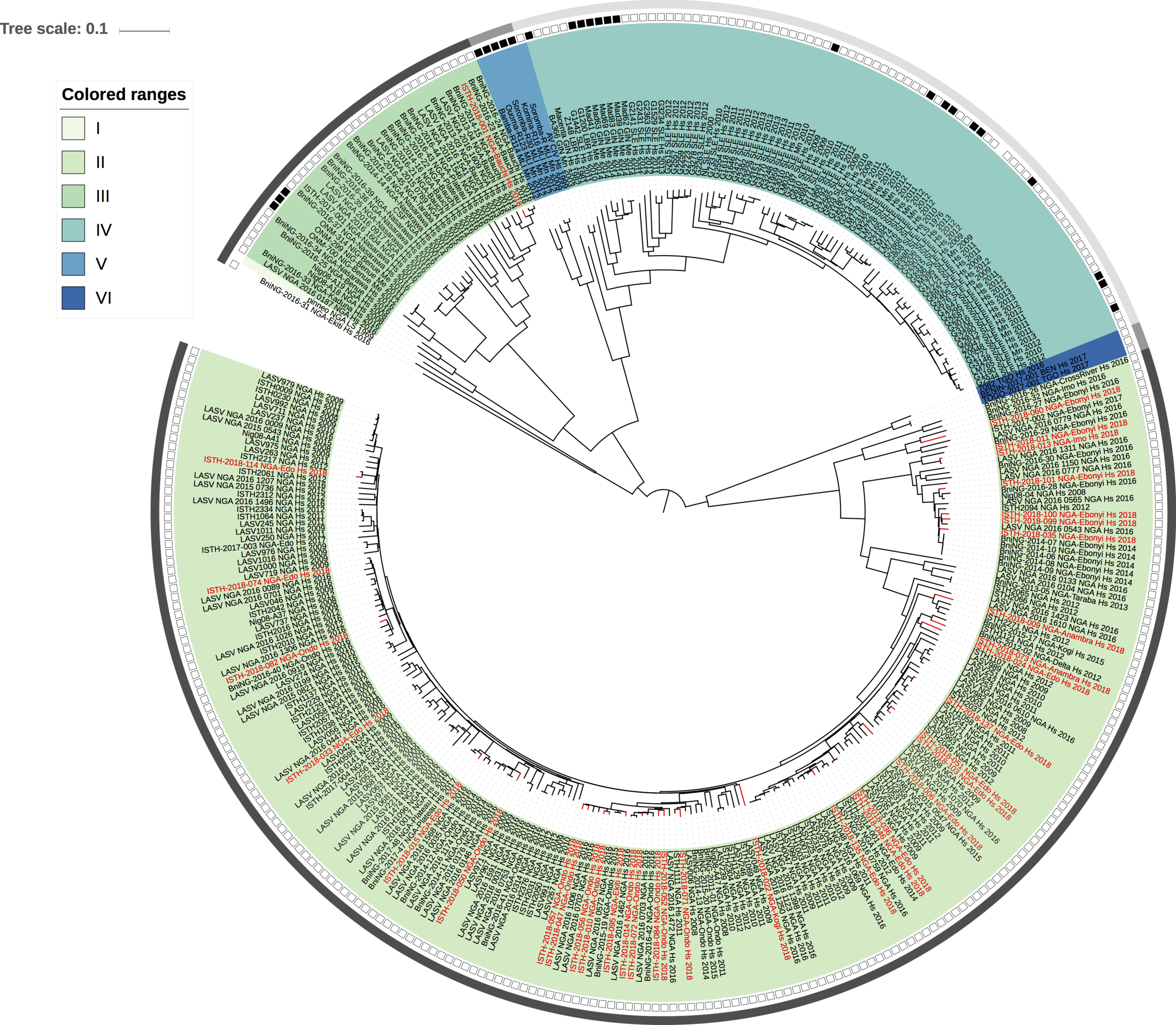
Maximum likelihood phylogenetic analyses of the coding genes in both the L and S segments indicates more spill-over from the rodent hosts, now also in lineage III. However, we start to observe clusters of pairs of 2018 viruses (ISTH-2018-010 & ISTH-2018-056, and ISTH-2018-011 & ISTH-2018-013). In order to estimate the time of their most recent common ancestors (MRCAs), we performed BEAST analysis on the relevant subset of the S segment data. Divergence times and evolutionary rates are consistent with those previously estimated by Andersen et al. (doi: 10.1016/j.cell.2015.07.020). The time estimates demonstrates that the MRCAs dates back to 2013 (2011-2015) for ISTH-2018-010 & ISTH-2018-056 and to 2015 (2013-2016) for ISTH-2018-011 & ISTH-2018-013. So, this makes human-to-human transmission also unlikely for these pairs, indicating that -as for the other viruses - they are the result from independent reservoir spill-over. As a more dense sampling will become available, we expect to see more clustering of 2018 strains.

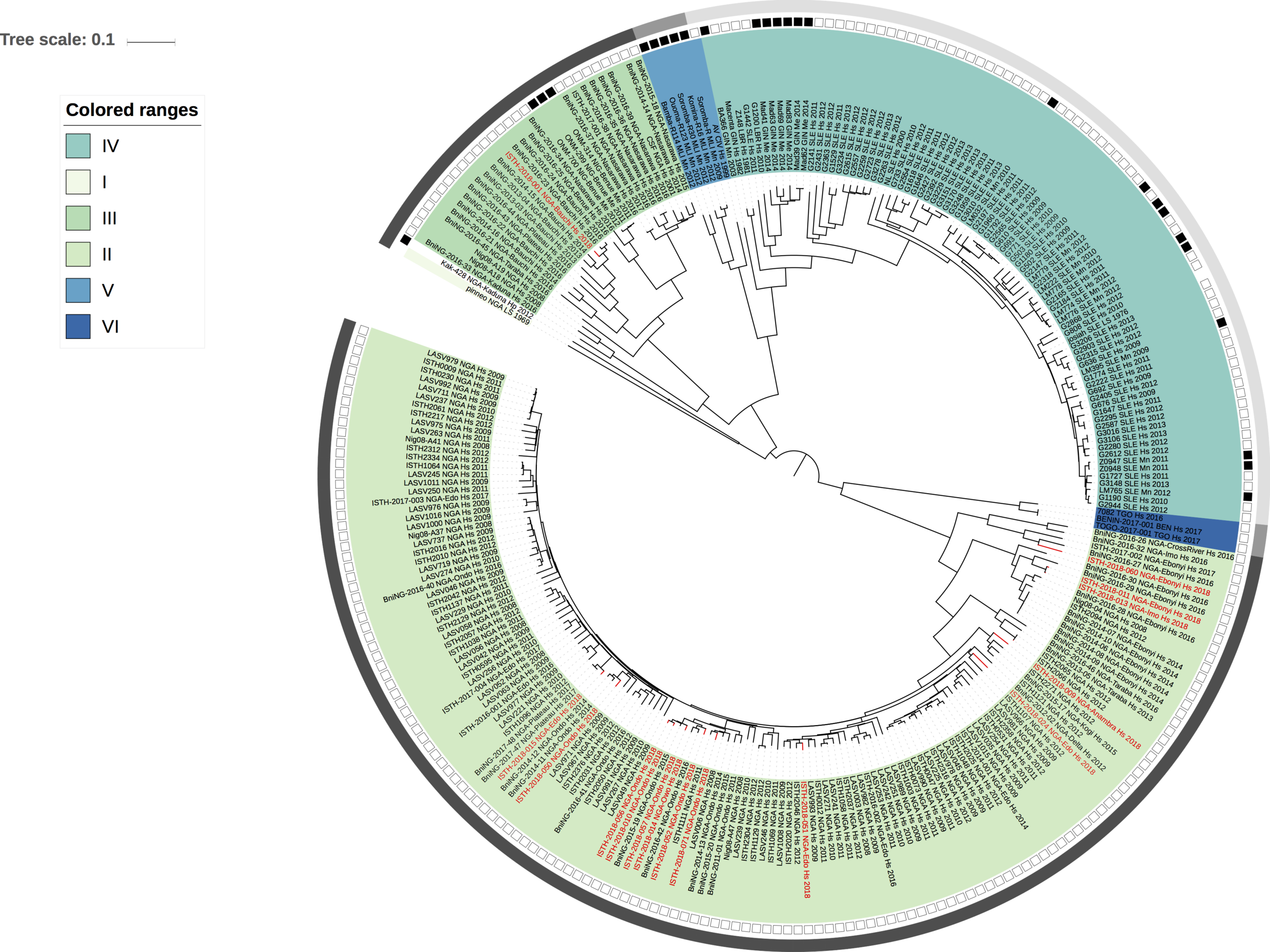
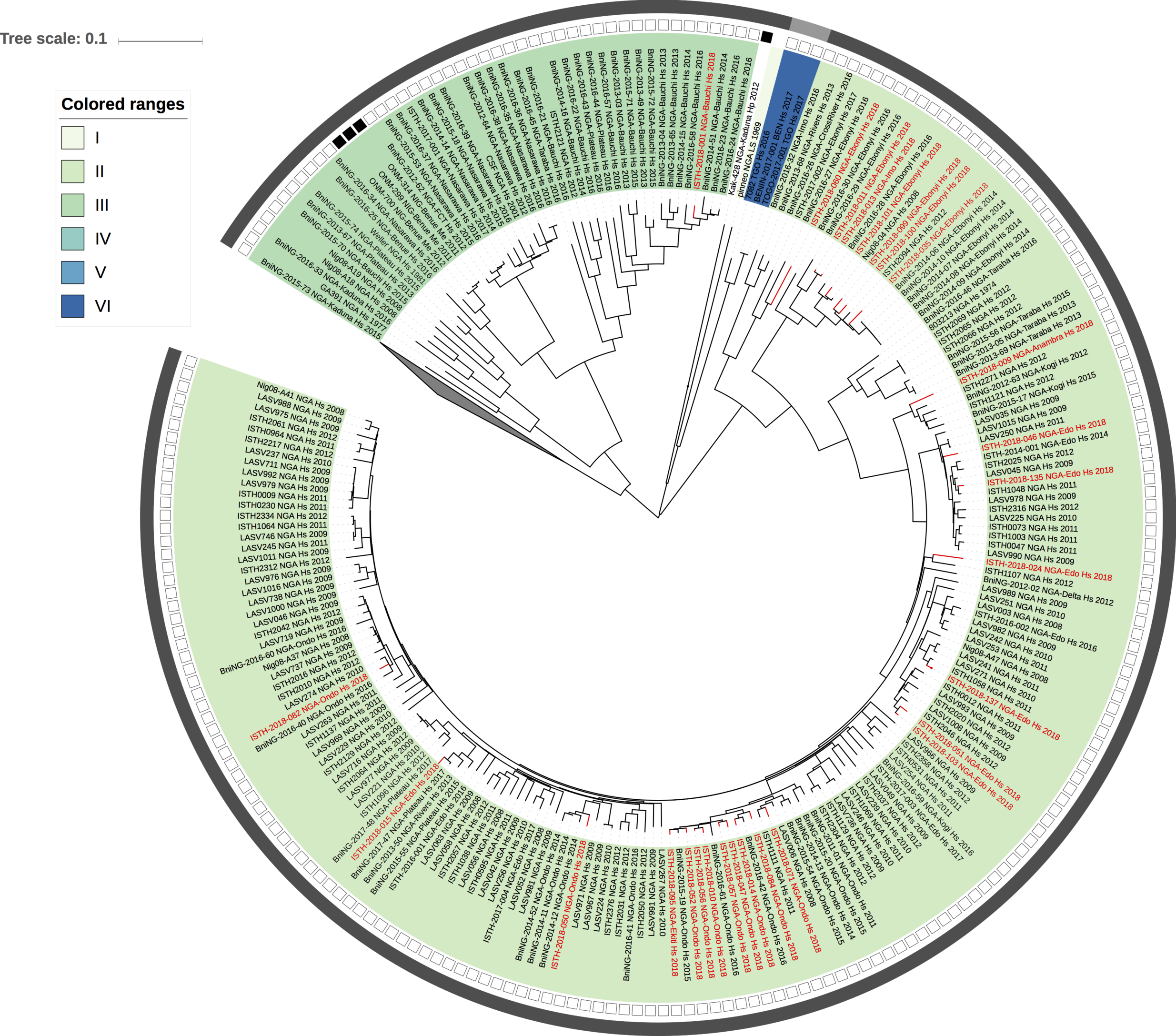
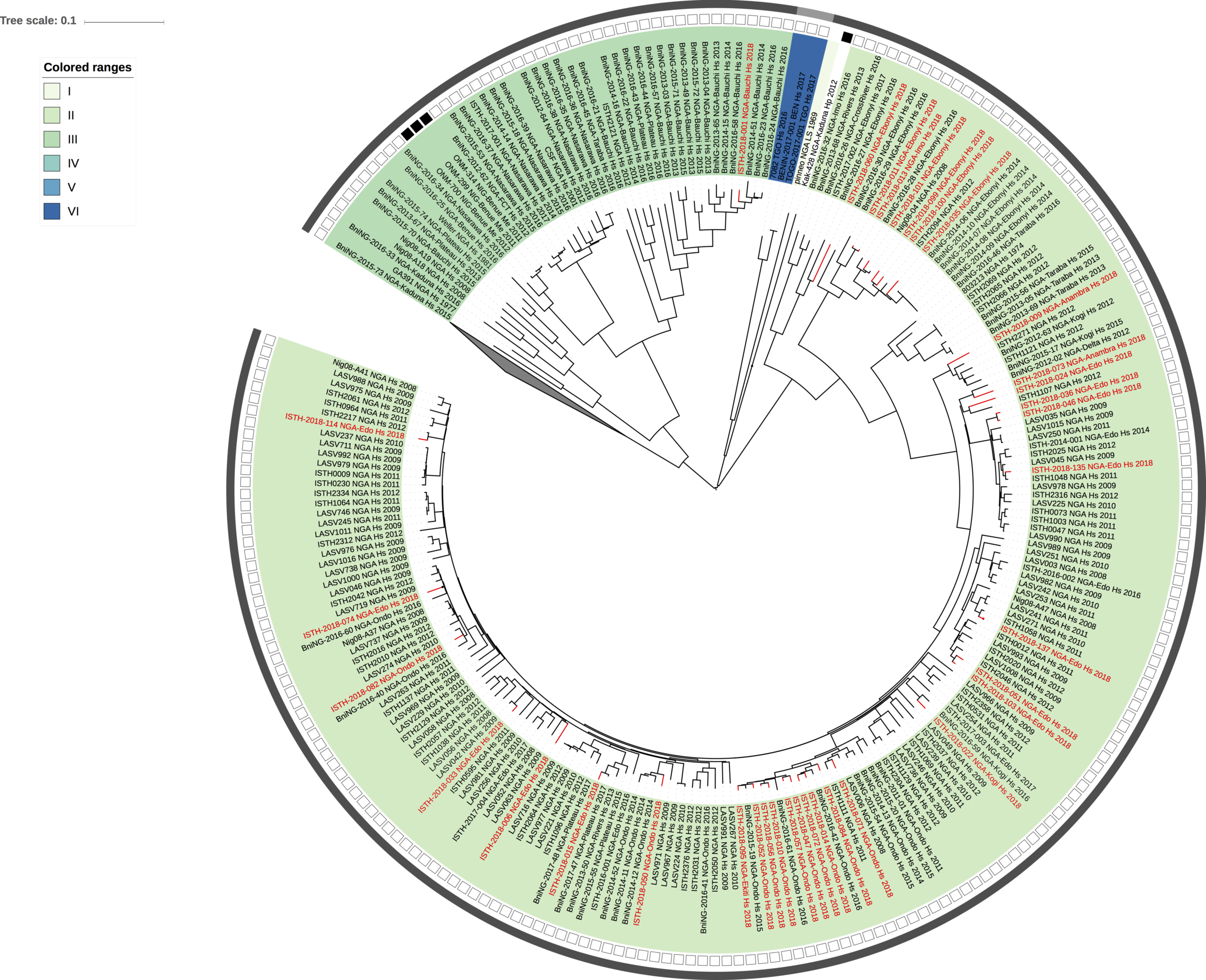
Figure 1: Lassa virus L-segment phylogeny. The circular tree includes all LASV lineages (I-VI) represented by different color ranges. The squares at the tips represent host: empty square = human virus; black square = rodent virus; no square = laboratory virus. The outer circle represents geographic origin: dark grey = Nigeria, light grey: Mano River Union countries, intermediate grey = other (Togo, Benin & Mali). Newly sequenced viruses are depicted in red.

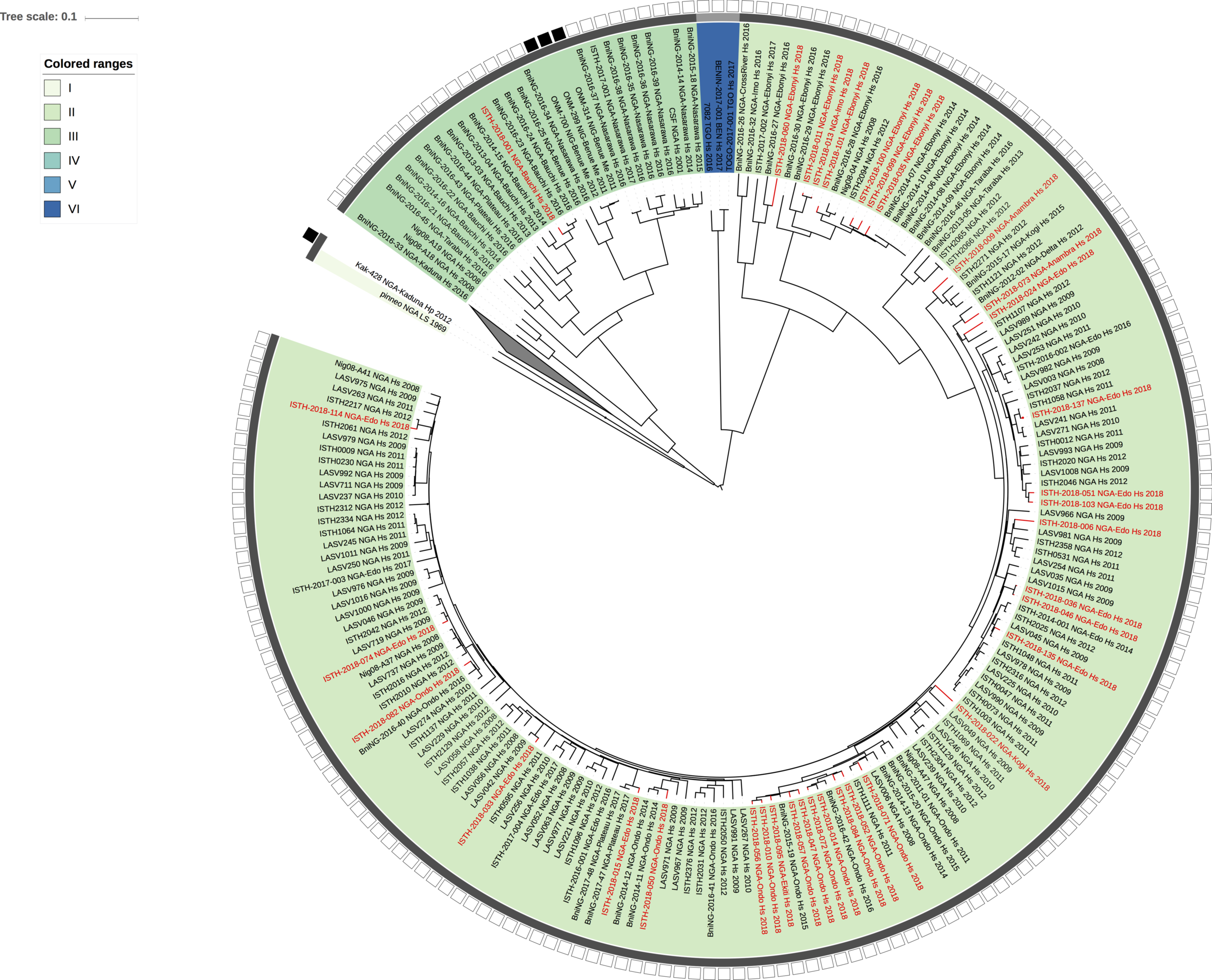
Figure 2: Lassa virus S-segment phylogeny and time-measured tree estimates. The circular tree includes all LASV lineages (I-VI) represented by different color ranges. The squares at the tips represent host: empty square = human virus; black square = rodent virus; no square = laboratory virus. The outer circle represents geographic origin: dark grey = Nigeria, light grey: Mano River Union countries, intermediate grey = other (Togo, Benin & Mali). Newly sequenced viruses are depicted in red. The time-measured tree includes all lineages except for lineage IV and V. Two clades are indicated that contain a pair of 2018 sequences. These clades and their timescale are shown in more detail on the right.

DISCLAIMER
The Irrua Specialist Teaching Hospital (ISTH), Edo State, Nigeria, Bernhard Nocht Institute for Tropical Medicine (BNITM), Hamburg, Germany, and Public Health England (PHE), UK, are committed to sharing data in public health emergencies. We release unpublished Lassa virus sequences from Nigeria to support the public health response as well as the development and evaluation of Lassa fever diagnostics or therapeutics. The data may be used and analyzed for these purposes. It is not permitted to use the sequences for publication, i.e. any type of communication with the general public that describes data generated with the use of the sequences. If you intend to so, please contact us directly.

Dr. Ephraim Epogbaini [email protected], Director of Institute of Lassa Fever Research and Control,

Prof. Stephan Günther [email protected], Director, WHO Collaborating Centre for Arboviruses and Hemorrhagic Fever Reference and Research, BNITM

Thanks Philippe for this analysis. Perhaps the most important thing to do with these sequences to better understand the current situation is to analyse them in concert with the 2015-2016 genomes recently posted on this site: New Lassa Virus genomes from Nigeria 2015-2016

The statements on the use of the data from the two groups might preclude such an analysis until the respective publications are out? Perhaps something could be worked out?

Thanks Philippe. Great observations here. In our 2015 paper we saw a couple of clusters (<5% of all sequences) that could have been the result of human-to-human transmissions. In those cases, the sequences were either completely identical, or only had 1-2 SNPs between them. While such a picture is consistent with human transmission, it could also be because people were infected in the same household, from the same rodent(s). Just something to keep in mind if clusters start showing up - could be human transmission, but could also be transmission from the same rodent(s). Given your analysis and the one from ACEGID/Broad, I agree there’s currently no evidence to support (an uptick in) human transmissions.

Would be useful to have more rodent sequences but looking at the tree there are a few Me sequences (ONM-314, 299 & 700 - at about 10 o’clock on the tree) that are very close even when separated by a year (although with just the year not the full date, this could just be spanning the new year).

Formal analysis and modelling required to answer this question.

We can now include sequences for 12 new samples, so amounting a total of 27 samples:

https://github.com/ISTH-BNITM-PHE/LASVsequencing

Because these new viruses also cluster within lineage II and III, the maximum likelihood trees are shown with a collapsed clade for lineage IV and V (the Mano River Union countries clade):

Figure 1: Lassa virus L-segment phylogeny. The circular tree includes all LASV lineages (I-VI), but the clade for lineage IV and V is collapsed for clarity. The squares at the tips represent host: empty square = human virus; black square = rodent virus; no square = laboratory virus. The outer circle represents geographic origin: dark grey = Nigeria, lighter grey = other (Togo, Benin & Mali). Newly sequenced viruses are depicted in red.

Figure 2: Lassa virus S-segment phylogeny. The circular tree includes all LASV lineages (I-VI), but the clade for lineage IV and V is collapsed for clarity. The squares at the tips represent host: empty square = human virus; black square = rodent virus; no square = laboratory virus. The outer circle represents geographic origin: dark grey = Nigeria, lighter grey = other (Togo, Benin & Mali). Newly sequenced viruses are depicted in red.

Because more 2018 sequences are clustering together, we have again performed a BEAST analysis for the S segment data in order to estimate times to the most recent common ancestor of each 2018 sequence with the most closely related sequence. This indicates that it takes considerable time, well over 1 year (red line in Figure 3) for all samples, to find that common ancestor, indicating that they are not directly linked by human-to-human transmission and also unlikely to be part of a human-to-human transmission chain. In conclusion, and in agreement with Kristian’s comment, there remains no evidence for human transmission in this 2018 data.

Figure 3: box plot of times to the most recent common ancestor of each of the 27 2018 sequences with their most closely related sequence as estimated using BEAST. The two sequences with the shortest time estimates, ISTH-2018-011 & ISTH-2018-013, for a cluster pair as shown in the analysis of the previous post.

Sequences have now been generated for 8 additional samples, so for 35 samples in total:

https://github.com/ISTH-BNITM-PHE/LASVsequencing

Also these 2018 viruses cluster within lineage II and III, so we collapsed the clades for lineage IV and V (the Mano River Union countries clade) in the maximum likelihood trees:

Figure 1: Lassa virus L-segment phylogeny. The circular tree includes all LASV lineages (I-VI), but the clade for lineage IV and V is collapsed for clarity. The squares at the tips represent host: empty square = human virus; black square = rodent virus; no square = laboratory virus. The outer circle represents geographic origin: dark grey = Nigeria, lighter grey = other (Togo, Benin & Mali). Newly sequenced viruses are depicted in red.

Figure 2: Lassa virus S-segment phylogeny. The circular tree includes all LASV lineages (I-VI), but the clade for lineage IV and V is collapsed for clarity. The squares at the tips represent host: empty square = human virus; black square = rodent virus; no square = laboratory virus. The outer circle represents geographic origin: dark grey = Nigeria, lighter grey = other (Togo, Benin & Mali). Newly sequenced viruses are depicted in red.

ISTH-2018-011 & ISTH-2018-013 remain the two 2018 sequences with the most recent common ancestor, but as shown by the BEAST analysis in our previous post, also these are unlikely to be linked by human-to-human transmission.

The 35 2018 genomes have now been put together with unpublished Lassa virus sequences (partial and complete) from previous years, that are under analysis by BNITM, and with the 64 2015-2016 genomes recently posted by colleagues at http://virological.org/t/new-lassa-virus-genomes-from-nigeria-2015-2016/191.

11 duplicate genomes from the same samples have been removed. Maximum likelihood phylogenetic analyses of the coding genes in both the L and S segments confirm previous conclusions about the independent spill-over from the rodent reservoir.

Figure 1: Lassa virus S-segment phylogeny. The circular tree includes all LASV lineages (I-VI), but the clade for lineage IV and V is collapsed for clarity. The squares at the tips represent host: empty square = human virus; black square = rodent virus; no square = laboratory virus. The outer circle represents geographic origin: dark grey = Nigeria, lighter grey = other (Togo, Benin & Mali). Newly sequenced viruses are depicted in red.

Figure 2: Lassa virus L-segment phylogeny. The circular tree includes all LASV lineages (I-VI), but the clade for lineage IV and V is collapsed for clarity. The squares at the tips represent host: empty square = human virus; black square = rodent virus; no square = laboratory virus. The outer circle represents geographic origin: dark grey = Nigeria, lighter grey = other (Togo, Benin & Mali). Newly sequenced viruses are depicted in red.

Acknowledgements.
The Irrua specialist Teaching Hospital (ISTH), Institute for Lassa Fever Research and Control (ILFRC), Irrua, Edo State, Nigeria
The Bernhard-Nocht Institute for Tropical Medicine (BNITM), Hamburg, Germany,
Public Health England (PHE),
African Center of Excellence for Genomics of Infectious Disease (ACEGID Redeemer’s University, Ede, Nigeria),
Broad Institute of MIT and Harvard (Cambridge, MA, USA), Harvard University (Cambridge, MA, USA)